English

A urinalysis test can provide critical information in a way that is cost-effective and noninvasive. Urine is collected midstream or by catheterization and is examined within one hour after collection to avoid destruction of formed elements.1 Urinalysis typically then undergoes a chemical test, using reagent test strips, and microscopic evaluation. Additional tests may include microbiology, cytology and others.
This information can be used to screen for a wide range of disorders including kidney diseases, metabolic disorders, cancer, infections, etc. Various findings on the urinalysis can also indicate certain patterns of kidney disease and whether the disease is likely to be chronic versus acute in nature.
Acute kidney injury (AKI) is the deterioration of kidney function over hours or days, resulting in the retention of nitrogenous wastes and creatinine in the blood. Chronic kidney disease (CKD) results from the gradual loss of kidney function over months or years. Often, heavy proteinuria and lipuria are consistent with nephrotic syndrome.1
A common clinical approach in assessing kidney dysfunction is the cause and severity of the renal abnormalities. In all cases, this evaluation includes the patient history, physical exams, a urinalysis test, glomerular filtration rate (GFR) and urine albumin-creatinine ratio (ACR). Alongside a patient’s eGFR, urine tests can help give a more accurate picture of how well their kidneys are working.1
What is the Importance of Urine Microscopy?
Urine microscopy consists of the evaluation of particles such as WBC, RBC, casts, crystals, epithelial cells and more. These particles, more specifically casts and crystals, may provide evidence of significant renal disease in patients. Urine microscopy is vital to aiding in the diagnoses of many asymptomatic cases and for those that are unable to verbalize their symptoms to their physician, whether due to coma or otherwise. These include urinary tract infection (UTI), urinary tract tumors, kidney disease and nephrotic syndrome.2
Formation and Significance of Urinary Casts
Casts are cylindrical particles that clump together and formed either in the distal convoluted tubules or the collecting ducts of the kidney. The walls of the tubules act as a mold for cast formation and the width of the tubule determines the width of the cast. As a result, narrow casts are formed in the distal tubules while broad casts are formed in the collecting ducts.3
One example of a protein cast is the accumulation of the Tamm-Horsfall mucoprotein. This protein entraps cells and materials within the kidney tubes resulting in protein casts. The elements which favor protein cast formation are low flow rate, high salt concentration and low pH, resulting in protein denaturing and precipitation.4
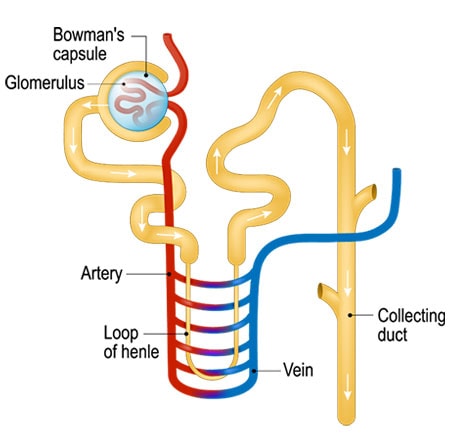
Figure 1: Renal tubules are metabolically active, being responsible for absorption or excretion of a wide range of substances.
Very few casts are seen in the urine of a person without renal disease. A common exception is hyaline casts, which may often be seen in healthy patients or can be present after strenuous exercise or diuretic use. A significant number of urinary casts usually indicates the presence of renal disease.3
Formation and Clinical Significance of Urine Crystals
Urine crystals form when urine has an excessive number of minerals present. Different types of urine crystals form for different reasons. Underlying health conditions, eating a diet too high in protein/salt or dehydration can cause urine crystals to form. Consequently, when there is an excessive buildup of one or more minerals, a urine crystal can form into a stone.5
Normally, urine crystals will have limited symptoms unless large enough stones develop. When this happens, the stone may pass naturally out of the body, or medical intervention may be required to help remove the stone.5
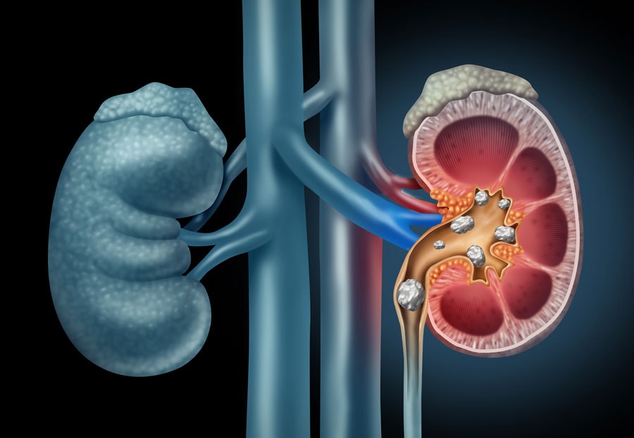
Figure 2. When there is an excessive buildup of one or more minerals, a urine crystal can form into a stone. A kidney stone usually will not cause symptoms until it moves around within your kidney or passes into your ureters. If it becomes lodged in the ureters, it may block the flow of urine and cause the kidney to swell and the ureter to spasm, which can be very painful
Early Detection is the Most Effective Way to Combat Kidney Disease
A quick, non-invasive urinalysis test highlights the importance of the laboratory’s role in patient care. At least 70% of all physician clinical decisions are made from laboratory results.6 The goal of any hospital or clinical lab is to provide the highest level of care to the patient, and the technology a lab utilizes plays a major role in achieving this purpose.
This is especially important with CKD as many patients have no idea that their kidneys are failing. Damaged kidneys leak protein into the urine when it should be in the bloodstream and it is the presence of casts and/or crystals on a urinalysis, that may be one of the first early indicators of underlying kidney disease.7
For more than 30 years, Iris urine analyzers have been providing laboratories automated urine microscopy to aid clinicians in the diagnosis of diseases such as kidney disease.
The iQ200 Series urine microscopy analyzer automatically identifies 12 particle classifications and 27 sub-classifications with >90% confidence.8 This automated urine analyzer has the most extensive classification capability on the market with the ability to create user-defined reports specific to laboratorians needs. Automated urine analyzers utilizing auto-particle classification and digital imaging provide laboratories the capacity for auto-quantitation and categorization of urine microscopy results.
See why the presence of urinary casts and urinary crystals may indicate underlying evidence of kidney dysfunction.